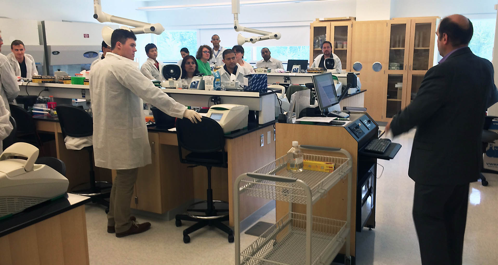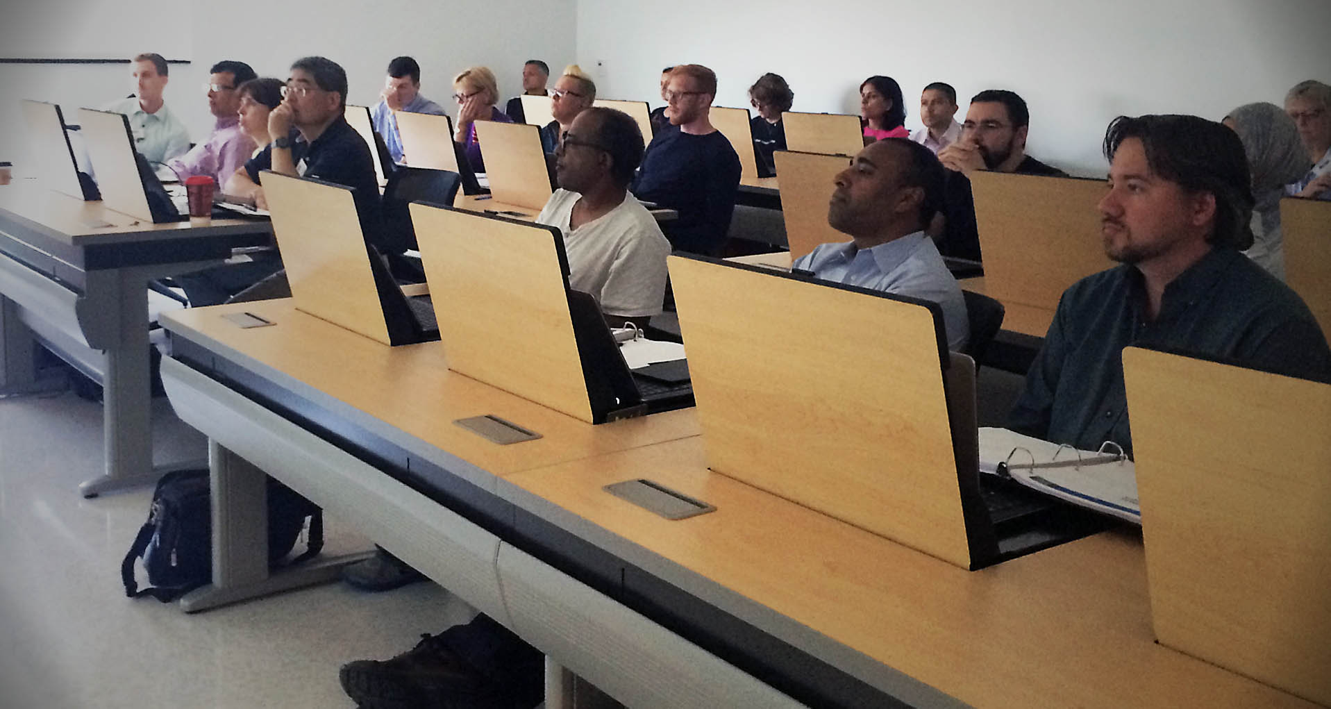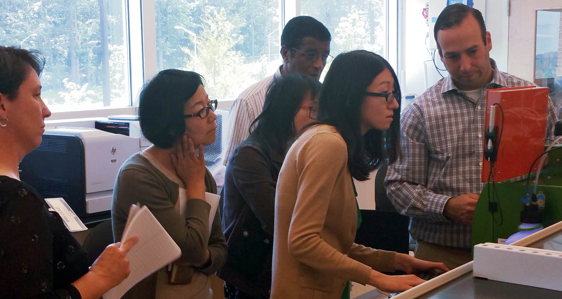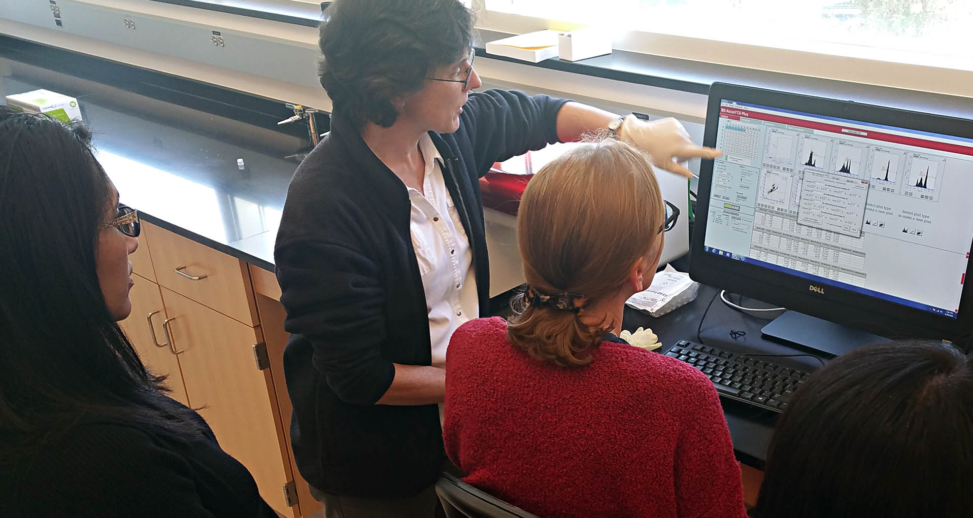

 | Lecture and Hands-on Interactive Training |
 | Team taught by active researchers |
 | Thumbnail drive with Lectures and Workshop material |
 | Space limited to 24 participants |
 | Registration Fee: $1,895.00 |
Course Directors

- Pre-analytics: Sample acquisition, fixation, processing embedding and microtomy
- Reagents and validation: Reagents, antibody validation, protocol optimization and controls
- Fundamentals of labels and stains: color, chromogen and fluorescent detection systems
- Microscopy basics: transmitted light and fluorescence microscopes, limitations of the microscope and slide
- Faculty facilitated discussions
- Troubleshooting sessions
- Advanced immunohistochemistry topics: antigen retrieval, multiplex immunohistochemistry, other detection systems and molecular targets
- Horizon Lectures – automated IHC, quantitative digital pathology, digital imaging and deep learning, highly multiplexed IHC and TBA
- Immunohistochemistry laboratory
- Dual label immunofluorescence laboratory
- Microscopy and digital imaging
- Coverslipping
 | A. Sally Davis DVM, PhD, DACVP, DACVM Resident Education and Outreach Lead, The Histochemical Society, Rockville, MD Adjunct Faculty, Department of Diagnostic Medicine/Pathobiology, Kansas State University, Manhattan, KS Extraordinary Faculty, Department of Paraclinical Sciences, Veterinary Faculty, University of Pretoria, Onderstepoort, Gauteng, South AfricaPhD Sally Davis is a board-certified veterinary pathologist and microbiologist whose research focuses on emerging and zoonotic viral pathogens. She has 15 years of experience in the development of tissue-based assays, particularly immunohistochemistry, for a diverse array of species, tissue and target types. She is an educator who has taught both trainee and established researchers these techniques in her laboratory at Kansas State University. |
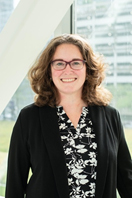 | Francesca E. Duncan, PhD Assistant Professor, Department of Obstetrics and Gynecology, Feinberg School of Medicine, Northwestern University Co-Director, Center for Reproductive Science, Northwestern University Assistant Professor-in-Residence, Buck Institute for Research on Aging, Novato, CA Francesca Duncan is a reproductive biologist whose research focuses on mechanisms of female reproductive aging. Given the complex tissue architecture and cellular heterogeneity of the mammalian ovary, histology and histochemical approaches are central to her research in multiple species including human, nonhuman primate, cow, pig, mouse, and naked mole rat. She is a proud alumna of the 2016 Immunhistochemical and Microscopy course which is the predecessor of the current course and now serves on the Histochemical Society Council. |
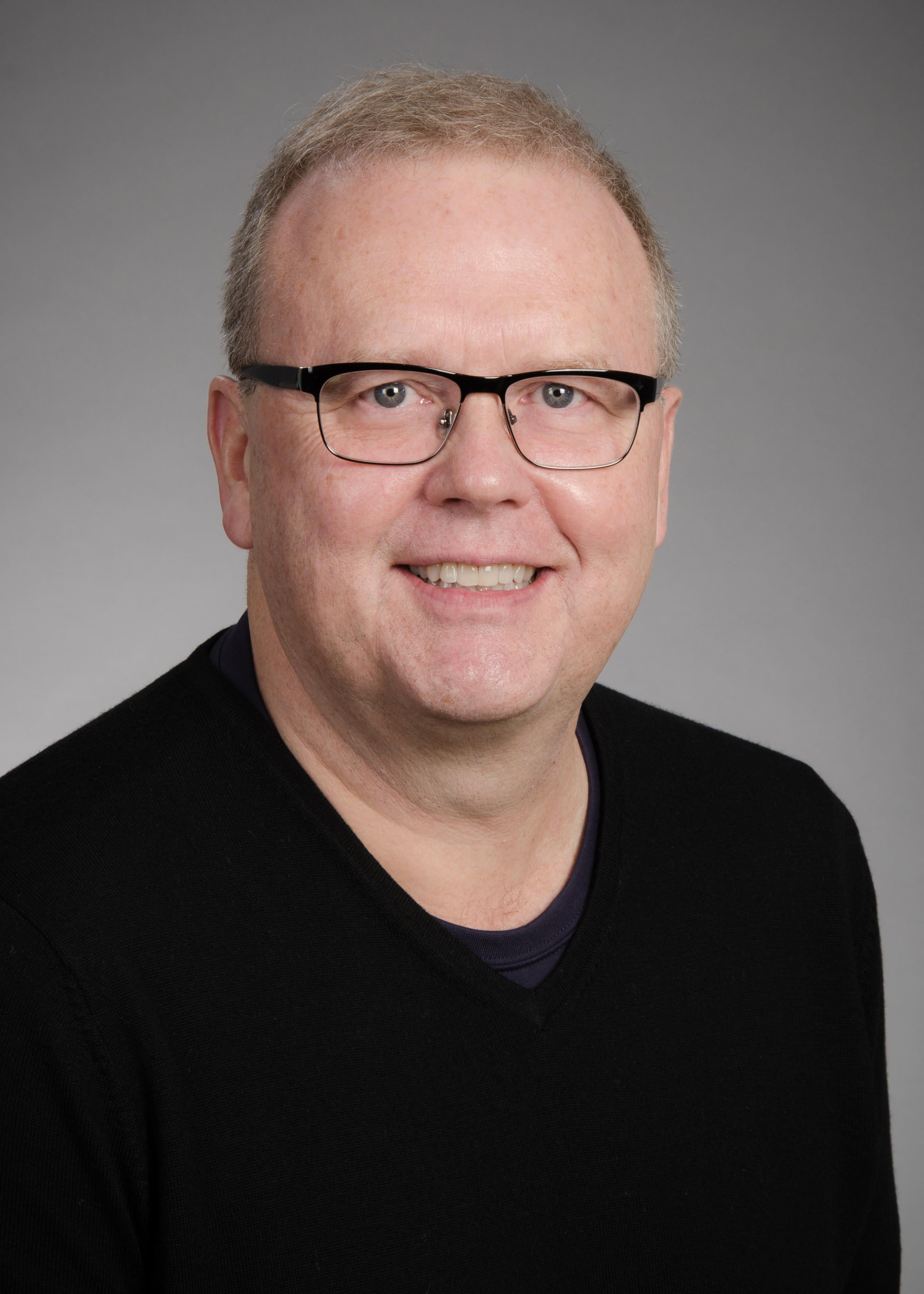 | Charles W. Frevert, DVM, ScD Professor, Department of Comparative Medicine, University of Washington (UW), Seattle, WA Professor, Department of Medicine, Division of Pulmonary Critical Care Medicine and Sleep Medicine, UW Adjunct Professor, Department of Pathology, UW Charles Frevert, a veterinary scientist, and comparative pathologist, is a professor in the Department of Comparative Medicine at the University of Washington School of Medicine. In addition to his research, Dr. Frevert is the Director of the Histology and Imaging Core, a state-of-the-art research pathology laboratory at the University of Washington specializing in immunohistochemistry and quantitative digital pathology. |
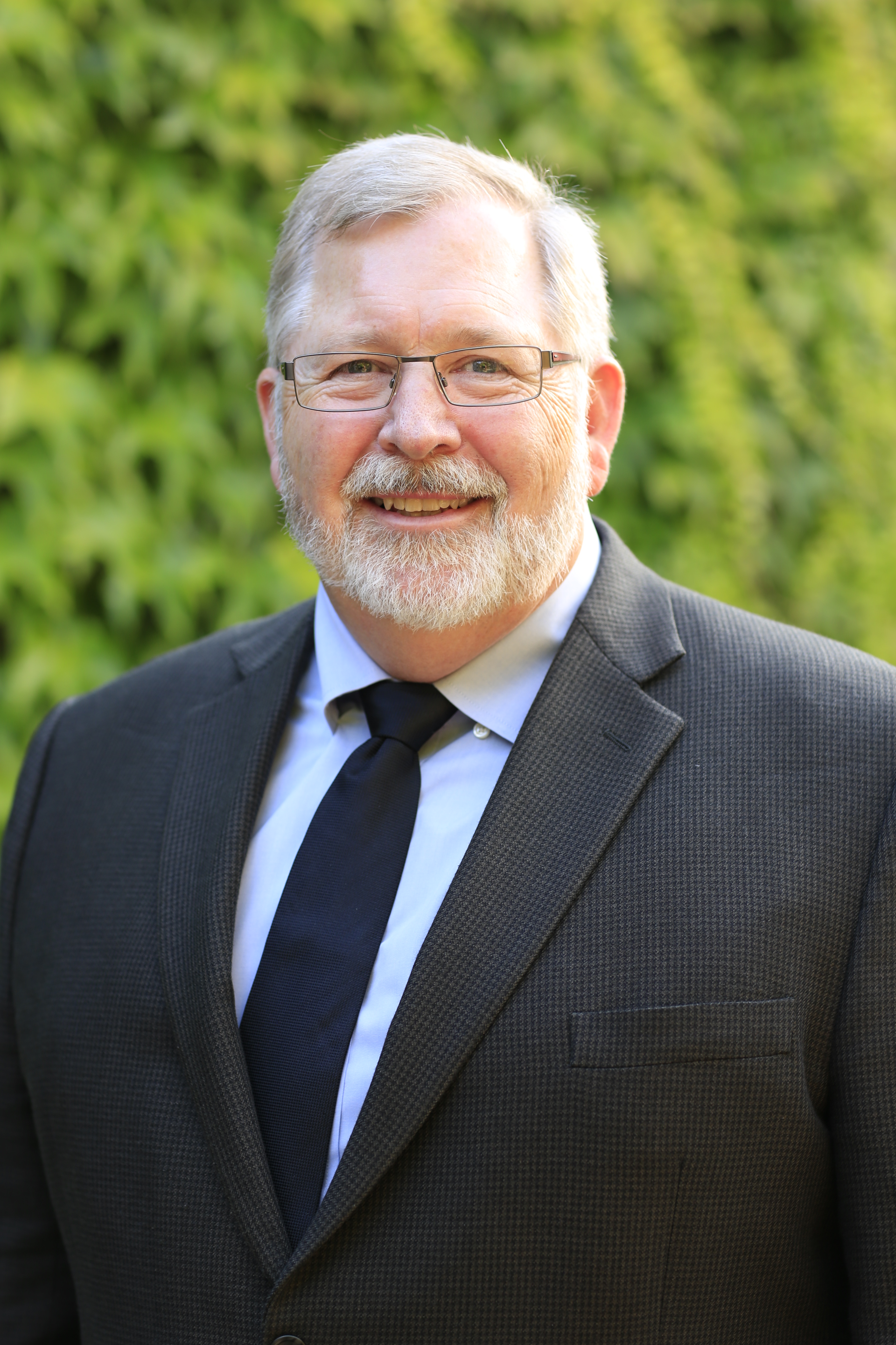 | Paul C. Goodwin, MS Past-President, The Histochemical Society Science Director, Cytiva, Issaquah, WA Affiliate Teaching Associate, Department of Comparative Medicine, University of Washington, Seattle, WA Paul Goodwin is the Science Director for Cytiva and resides in Seattle, WA. He explores the future of science, technology, and business models that will affect Life Sciences and investigate ways to convert challenges into business opportunities. He has been on the faculty for numerous microscopy courses at the Marine Biological Laboratory (MBL) in Woods Hole, MA, and Cold Spring Harbor Laboratories, Cold Spring Harbor, NY, for many years and he is the Past-President of The Histochemical Society. |
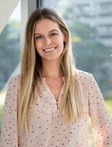 | Madison Gowett Graduate Student, Department of Obstetrics and Gynecology, Feinberg School of Medicine, Northwestern University Madison Gowett is a master’s student at Northwestern University in the Duncan Lab and her research focuses on ovarian aging. Specifically, she is interested in developing novel 3D culture approaches to study aging in the ovarian stromal compartment, and she utilizes various histochemical techniques to characterize this system. |
 | Stephen M. Hewitt. MD, PhD Head, Experimental Pathology Laboratory, Center for Cancer Research, National Cancer Institute, NIH, Bethesda, MD Stephen Hewitt is Editor-in-Chief of the Journal of Histochemistry & Cytochemistry and co-chaired the Clinical Laboratory Standards Institute committee on guidelines for immunohistochemistry in clinical diagnostic laboratories. |
 | Frank Peale Distinguished Pathologist, Genentech, South San Francisco, CA Frank Peale has 24 years’ experience in Genentech’s Research Pathology department collaborating with other investigators to provide tissue-based endpoints, including immunohistochemistry, for basic science and early clinical development projects. He has a particular interest in developing methods to allow antibody-mediated staining to be performed and evaluated consistently in different labs. |
 | This year we welcome Andrea J. Radtke, PhD National Institute of Allergy and Infectious Diseases (NIAID.) Dr. Radtke will give a horizon lecture on Multiplexing methods and antibody validation for large scale tissue mapping initiatives. Dr. Radtke is an Associate Scientist at the National Institutes of Health. Andrea specializes in advanced microscopy techniques including a multiplexed antibody-based imaging method, IBEX, she developed with colleagues. Andrea is passionate about team science and leads several community efforts within the field of spatial biology. |
 | Leon Schermerhorn, DVM Leon Schermerhorn is a licensed small animal veterinarian with a special interest in veterinary anatomic and experimental pathology. He has worked with Sally Davis on various projects including IHC protocol automation and primary cell culture of zoonotic mammalian pathogens. He particularly enjoys immunohistochemistry and exploring its uses in diagnostic and experimental settings. |
 | Scott Tanner, PhD Assistant Professor of Biology, Division of Natural Sciences & Engineering University of South Carolina Upstate Scott Tanner is an Assistant Professor at the University of South Carolina Upstate, where he focuses on undergraduate education. He was trained in the pathology laboratory of Dr. Robin Lorenz. Using C. elegans, his current research focuses on development of the intestinal barrier. He has experience using immunohistochemistry in a variety of tissue types. |
June 2022
“I really enjoyed the workshop. All the teachers are so knowledgeable and patient. I can feel their intelligence, wisdom and passion. I hope I can be able to join the workshop again if my boss can send me here.” Jiong L., USUHS
“This was a great course to scale-up techniques in your lab. A great opportunity to interact with experts in the field.” Mar H., Texas State University
“This was an amazing course to learn from experts in the field. The hands on training and theoretical understandings and lectures was very informative! Additionally, I was able to build my network and collaboration list.” Letiticia M., ThermoFisher Scientific
“I loved this course! The knowledge that I gained from all the course instructors & participants will have an immense impact on my work, my images are going to look amazing!” Glennys R., NIH/NIAID
“This workshop gave me a good foundation on applying IHC & IF techniques. To me this will be useful to have a better understanding in the mechanisms and better to troubleshoot. This will also help as I move towards automated systems.” Editih V., Gilead Sciences
“Qualify presentations from experts in their fields.” Brad R, USUHS
“This was a great opportunity to get hands on training!” Sabrina R., NIH/NCI
“Satisfaction.” Babajide A., Ajayi Crowther University
“Good program, topics, useful details.” Anya F, USUHS
“It was an intense but well-organized course for people to learn IHC & IF. I definitely will recommend this course to others! And we were well-fed by Kendra!! Thank you for everyone who created this course.” Shuhan Z., Albany Medical College
“This class has changed the game for me with Immunohistochemistry! I am looking forward to taking more Bio-Trac courses!” Adaku U., Wright State University
“A fantastic experience with top-notch facilities/ equipment with even better instructors. I would recommend this course to anyone at any level working in Histology.” Dylan L., Kansas State University
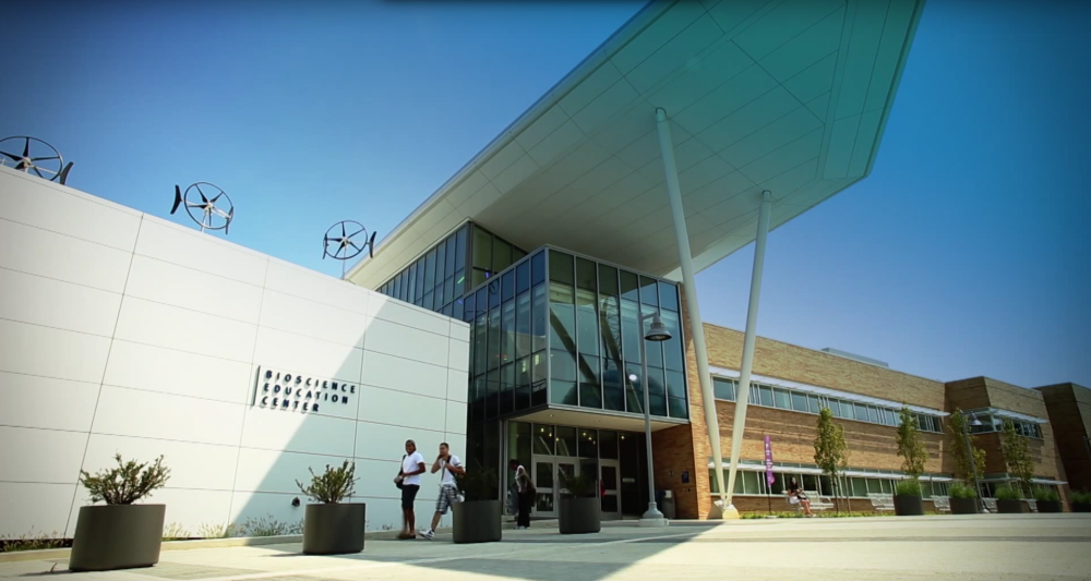
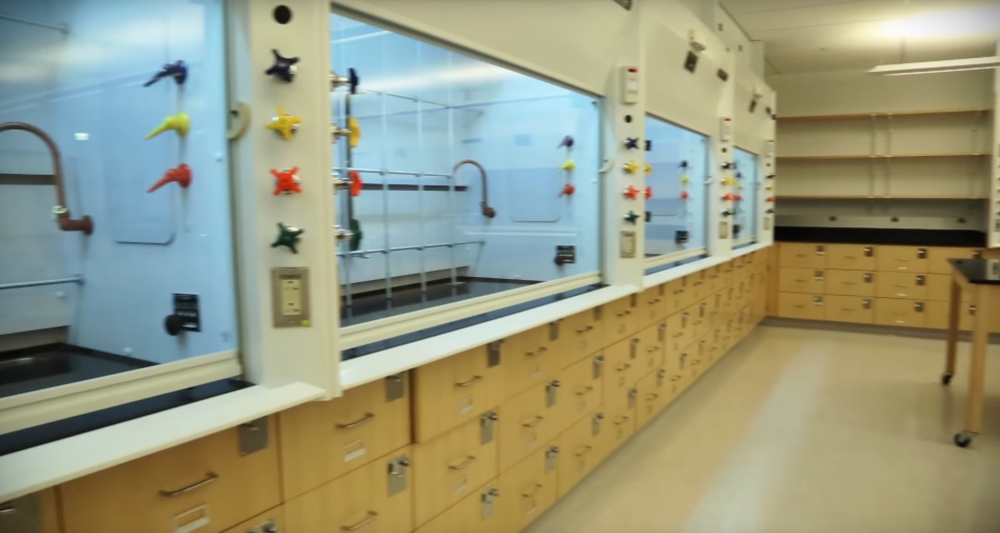
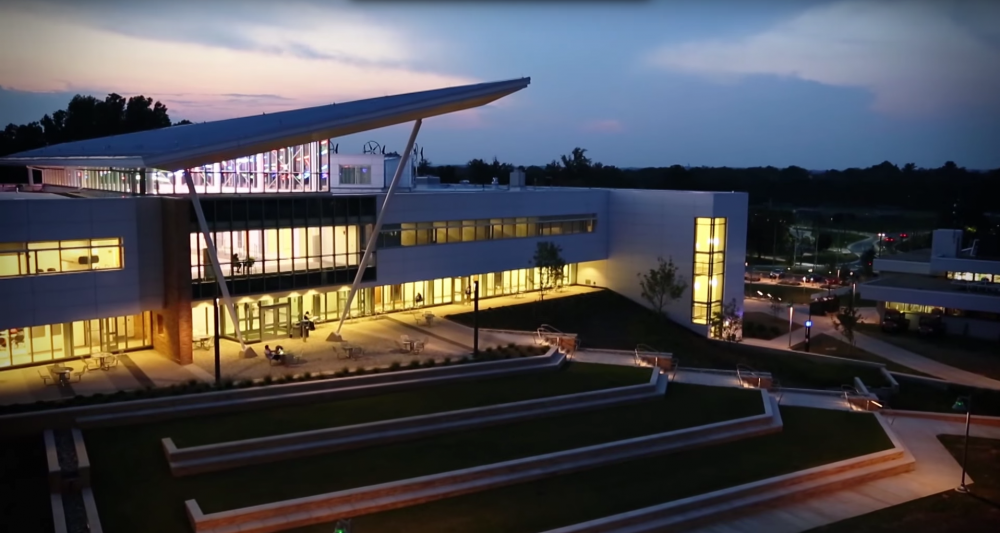
“The BEC facility is excellent! The lab spaces and technology were paramount to the course’s success. I enjoyed interfacing with Mark Nardone the course instructor. He kept students well-informed at all times.” Kelsey P, Kansas State University